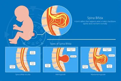
Spina bifida is a neural defect that occurs during fetal development resulting from a defective fusion of one or more posterior vertebral arches causing protrusion of the contents of the spinal canal before birth, incomplete closing of the embryonic neural tube (a group of cells that form the brain and the spinal cord of a baby). This can cause physical and mental issues. The neural tube and spinal column do not develop properly and remain exposed along several vertebrae. The spinal cord and membranes are pushed out forming a sac on the back of the fetus. This sac may be covered with membranes or meninges.
Types of spina bifida:
- Spina bifida occulta
- Spina bifida cystic(menifesta)
- Meningocele
- Myelomeningocele
Spina bifida occulta: There is merely a radiological defect between the laminae. Skin defects like haemangioma, lipoma, a tuft of hair, occur as a result of spina bifida occulta.
Spina bifida cystic: There is a developmental deficiency of laminae, spinous processes, overlying muscles, and skin.
Meningocele: In this condition mostly the spinal cord and its nerve roots are intact, only the covering membrane projects as a dural sac.
Myelomeningocele: It is associated with muscle paralysis, sensory loss, bladder and bowel incontinence, and deformities. It may be present from the 4th thoracic to 1st sacral vertebra. However, most common in the lumbosacral region. Associated with complications like paraplegia, deformities, bladder and kidney infections, skin ulceration, development of pathological fractures, meningitis as well as hydrocephalus.